Home Blog Page 97

The A, B, C’s of Oral Health

0
The A, B, C's of Oral Health

Food IS Medicine

What if we, in the dental profession, are doing dental healthcare all wrong? We continue to scale, polish, and lecture about oral hygiene, and our patients do their best to comply. Yet, dental disease is still at epidemic levels with at least 40% of the population having gum disease. By age 34, 80% of folks have had at least one cavity.

“Insanity is doing the same thing over and over, and expecting different results.” ~~~~~~~~~~~~~~~~~~~~~~~~~~~~~~~~~~~~~~~~~~~~~~Rita Mae Brown

I had a patient that had uncontrolled bone loss, red, bleeding tissues, and was losing teeth. She would see me every three months and I would give her my best recommendations, to no avail. Suddenly, one appointment she completely surprised me. Her gums were completely different- pink, tight, and little bleeding. She had completely changed her diet. She no longer ate carbohydrates and sugar, and it made all the difference for her oral health. That was an “ah-ha moment” for me. I knew I needed to look at dental health differently.

Maybe instead of working so hard to remove all the plaque and tartar during our dental hygiene recare appointments, instead we looked at the food fueling our cells, and investigate how to have a healthy microbiome. Most of our food is devoid of the nutrients our bodies need. Our soil is overfarmed, and our foods are full of chemicals. Too much of our food is over-processed and ultra-processed. Did you know the color additive ingredient titanium dioxide (or silicone dioxide or aluminum dioxide) contains lead, arsenic, and mercury? How can our bodies be healthy when our foods are full of chemicals that we know are hurtful to our cells?

Our bodies need good fuel for our cells.

Convience foods are high in sugar, salt and chemicals

Today’s post reviews the nutrients our mouth and teeth need to be healthy.

Vitamin A: a fat-soluble vitamin, it builds and maintains healthy teeth, bones, and oral tissues. It is also important for vision, reproduction, and fetal development.

A deficiency in Vitamin. A results in tooth decay, and dry mouth, dry eyes, dry skin.

Foods: fermented cod liver oil. (CLO is has the perfect ratio of Vitamin A to Vitamin D), greens, and orange and yellow vegetables.

B Complex Vitamins: Water soluble, they are essential for healthy gums. They accelerate gum healing.

Deficiencies in B’s result in mouth sores, dry mouth, cracked lips, bleeding gums, bad breath, angular cheilitis, and geographic tongue.

Foods with B’s: chicken, salmon, eggs, turkey, clams, mussels, green vegetables, and organ meats.

Vitamin C: is important for wound repair, and is an antioxidant. It helps the body to form and maintain connective tissues, including collagen found in gums and teeth. It prevents gum disease, builds strong enamel, and improves bad breath.

Deficiencies in Vitamin C: Scurvy, bleeding gums, easy bruising, poor wound healing, impaired immunity, swollen and painful joints, decrease in total collagen production and compromised collagen composition, and dry scaly skin.

Foods with C: Citrus, bell peppers, brussel sprounts, kiwi, kale, spinach, tomatos, and papaya.

CoQ10: Decreases gum inflammation, good for heart health, brain and muscles, and is important for growth and maintenance of the body. Helps heal our gums after periodontal therapy. It fights bacteria in the mouth, decreases gum inflammation, and helps maintain healthy teeth.

Deficiencies: Bad breath, tooth decay, dry mouth, and gum disease. Physical fatigue, mental fatigue, tired on waking, memory confusion, and difficulty concentrating. Most of us are deficient.

Foods with CoQ10: Fatty fish, organ meats, beef, sardines, and peanuts.

Collagen: Strengthens enamel, improves wound healing, gum health, and is essential for maintaining and repairing connective tissue.

Deficiencies: brittle teeth, receding gums, and mouth sores.

Foods: Sardines, organ meats, gelatin, eggs, beef, chicken with skin, bone broth.

Curcumin: an antioxidant and decreases gum inflammation and bleeding.

Foods: tumeric

Vitamin D3: Is important for gum health, and tooth health. Technically it is a hormone .It works with Vitamin A , K2 and Magnesium to feed the odontoblasts within the tooth and prevents tooth decay. It is important for bone health, sleep, and immune system.

Deficiency: tooth decay, increase in gum disease

Foods: Vitamin D is hard to get from the diet. Ideally, we get it from the sun, especially morning sunshine. Animal liver, fatty fish, egg yolks, and fish oils.

Green Tea: Has antibacterial activity, it helps in healing gums, tooth remineralization, and enamel health. It inhibits virus growth and decreases periodontal disease. It is proven to impede tumor cell growth.

Iodine: A trace mineral, it helps the body absorb calcium and promotes arterial and bone health. It is essential for a healthy thyroid gland.

Deficiency: increase in tooth decay, and gum disease, swollen tongue, dry mouth and mouth breathing.

Food: Sea salt, cod, spinach, bananas, cheese, lobster, eggs, and broccoli

Vitamin K2: Tells the calcium in the blood where to go- into the teeth and into the bones. It works with Vitamins D3 and A to help teeth heal. It improves insulin sensitivity which helps reduce gum inflammation.

Deficiency: Tooth decay, tartar build up, and athersclerosis.

Foods: Eggs, grass-feed butter, beef liver, natto, eel, fermented foods, sauerkraut, and chicken.

L-Arginine: An amino acid that is a biofilm manager, It buffers the pH of the biofilm and reduces biofilm thickness and density. The proper thickness of the biofilm helps protect the teeth. It helps remineralize teeth and reverses and prevents tooth decay. Saliva supplemented with arginine resits the lowering of pH, reducing the risk of demineralization of tooth surfaces.

Deficiency: Poor wound healing, diminished insulin production, atherosclerosis, high blood pressure, skin rash and loss of hair.

Food: Meats, fish, nuts, seeds, and legumes.

Magnesium: Mg works with Vitamins A, D3, and K2 for bone health and tooth health. Hardens tooth enamel, prevents loss of tooth density. It feeds the mitochondria.

Deficiency: Tooth decay, and dry mouth, restless sleep and restless leg syndrome. If you crave chocolate, you have a magnesium deficency.

Foods: chocolate, avocados, bananas, spinach , peanuts, almonds and kefir.

Melatonin: Stimulates bone cell that create bone and inhibits bone cells that break down bone which improves bone density. It promotes tissue regeneration. It promotes sleep. It helps protect the mitochondria.

Deficiency: Insomnia, depression, weight gain, acceleration of aging, metabolic disorders, and immunological aging.

Nitric Oxide: A gas produced by almost every cell in the body. It modulates the microbiome and reduces levels of anaerobic bacteria that cause bad breath. Nitric oxide is critical for brain and heart health. It improves our immune function. It makes the immune system resistant to viruses and bacteria. It may be the fountain of youth.

Deficiency: It reduces as we age and when we are not producing enough it ages you.

Food: Leafy greens, beets, garlic, meat, dark chocolate, citrus and pomogranate

Omega 3s: Improves connective tissue attachment and gum health and has antiinflammatory properties. Diets high in omega3s reduces t herisk of periodontal disease.

Deficiency: Depression, sensitive skin, dry skin, sun sensitivity, dry eyes, joint pain and stiffness, and hair changes.

Foods: fatty fish, flax seeds, chia seeds, and walnuts.

Potassium: Mineral that is important for the proper function of the heart, muscles, and nerves. It controls and maintains a healthy pH in the mouth which reduces the risk of tooth decay, periodontal disease and bad breath. It improves the mineral density of the teeth.

Deficiency: fatigue, confusion, muscle cramps and dry mouth.

Foods: spinach, tomatos, oranges, and bananas

Xylitol: a sugar alcohol that suppresses tooth decay bacteria strep mutans by making it less sticky, restores pH, prevents acidic bacteria from developing plaque, increases saliva flow, and keeps the teeth cleaner.

Zinc: forms and maintains the structure of the teeth gums and mucus membranes, and aids in saliva production. It is a mineral that is essential for the immune system, wound healing, normal taste and smell, prevents cavities, reduces inflammation, and is essential for healthy teeth and gums.

Deficiency: Dry mouth, cracked lips, hair loss, loss of taste and smell, tooth decay and gum disease, geographic tongue.

Foods: Oysters, beef, wheat germ spinach, mushrooms, yogurt and pumpkin seeds.

~~~~~~~~~~~~~~~~~~~~~~~~~~~~~~~~~~~~~~~~~~~~~~~~~~

Eat the rainbow at every meal

Food is medicine and we must look to it to help us be healthy and heal our bodies. We cannot supplement our way out of a poor diet. Garbage in=garbage out.

Supplements give our bodies a fighting chance. These foods, vitamins and minerals can help us have a healthy foundation and feed the good microbiome. We need to help the good bacteria, viruses and other microbes to grow and support us rather than allow the pathogens to take over. Before supplementing, please work with your primary care doctor to supplement properly for your body’s needs.

This list does not include every food, mineral or vitamin important to the mouth but it’s a great start. We also need fermented foods, water, as well as good sleep, stress reduction, and exercise. Our mouths would be healthier if we stopped eating sugar and fermentable carbohydrates.

Let’s conquer dental disease by looking at things differently. It’s time we got to the root of the matter- the food we ingest, the nutrients we need to have the building blocks for a healthy body. Take care of yourself and your loved ones by looking deeper into the foods you eat.

Barbara Tritz

Queen of Dental Hygiene and Life long Learner and Frustrated cook

Related

Myths About Root Canal Treatment

0
myths about root canal

A root canal can cause feelings of anxiety and fear. Root canals have the stigma of being painful and time-consuming procedures that can be very uncomfortable to endure. Some of the misconceptions about root canals are due to a lack of sufficient and correct information. Some others are because of the unpleasant experience of people with root canal treatment due to referring to non-specialist dentists, which has caused many people to avoid this common dental procedure. Although the treatment aims to treat your pain not cause it, there are many other myths about root canal that have made people afraid of it.

8 common myths about root canal

Here we will discuss common myths about root canal so that you won’t have any concerns.

1-Root canal is painful

Pulp infection and tooth inflammation cause pain and discomfort to the patient. With root canal treatment, this infection and inflammation are completely eliminated, and the toothache is relieved. Therefore, we can say that root canal not only will not cause pain, but it can also eliminate your tooth pain.

Today, dentists use local anesthesia and the patient will no longer feel any pain. In cases where the tooth infection is very severe, the patient feels a lot of pain, so the dentist prescribes antibiotics before the root canal treatment to reduce the pain and infection of the patient’s tooth.

2-Tooth Extraction is much better than root canal

Dentists are trying to preserve the patient’s natural teeth as much as possible. Tooth extraction creates an empty space in the mouth and causes complications. Among these complications, we can mention difficulty in chewing, jaw pain, bite problems, and displacement of adjacent teeth towards the empty space of the jaw.

As a result, root canal can be the best and most cost-effective way to save and preserve your natural teeth.

3-Root canal causes other diseases

Some people have the wrong idea that root canals will cause other diseases. Such a claim is completely wrong and root canal treatment is a safe and completely safe method. In addition, nerve removal causes the treatment of tooth infection and the destruction of bacteria in the root canal.

4-The tooth pain disappears immediately after the procedure

One of the misconceptions about root canal is the immediate disappearance of pain after the treatment. With denervation of the tooth, the nerve is completely destroyed and the pain signal is no longer transmitted. However, due to the inflammation of the adjacent tissues, the patient will feel pain in these areas for several days. According to the condition of the patient, this pain can be felt permanently or only during chewing.

Tooth pain after root canal is tolerable and the patient can take painkillers to relieve this pain until recovery.

5-Root canal means pulling the root of the tooth

Another myth about root canal is that, in this process, the root of the tooth is completely pulled. That seems beyond a myth.

In this procedure, the central part of the tooth called the pulp is cleaned and disinfected and it is filled with special materials. The root causes the strength of the tooth in the jaw and its stability, and if the root is removed, the tooth will fall.

6-You must visit the dentist for several sessions

The number of dental sessions in a root canal procedure depends on the number of treated teeth, complexity of root canals, and the level of tooth infection.

In cases where the tooth has a severe infection or you might need a crown, the procedure might take two dental appointments. At first, the dentist prescribes oral medicine and after a few days making sure the infection is completely removed, the treatment process can be completed. Typically, root canal treatment can be done in one hour to two hours.

7-If the tooth does not hurt, it does not need root canal

Pain and sensitivity are the most common symptoms of a pulp infection. When you delay a root canal to treat the damaged pulp, the infection gets worse and leads to pulp necrosis or pulp death.

As infections spread to the entire root canal, it causes dental abscess (formation of pocket of pus). You may have other symptoms like swelling, fever, or a bump near the tooth. This indicates a serious infection and you should visit the dentist as soon as possible.

If the tooth does not hurt, this doesn’t mean everything is right all the time. When the nerve dies, there is no signal of pain.

Also, the bump on your gum is actually a channel for the discharge of infectious secretions of the dental tissue. This channel takes the pressure resulting from the accumulation of infection inside the tissue and causes the patient not to feel pain in their tooth. This infection requires debridement and if it is not treated in time, it can cause infection of the adjacent tissues.

8-Root canal cannot be done on the same tooth again

Another false belief about root canal is if the tooth treated with this method becomes infected, you should pull it. Root canal treatment has a success rate of about 85%. But after a period if this tooth gets infected, your dentist or endodontist may opt for retreatment. Your tooth can be saved and preserved by repeating the process of root canal treatment.

How To Lose Belly Fat

0
Logo for WebMD

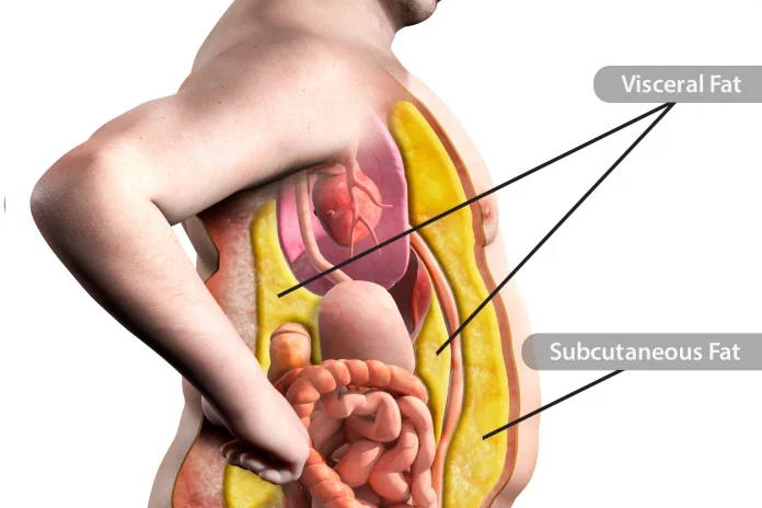
Surprise: Everyone has some belly fat, even people who have flat abs.That’s normal. Fat is an essential part of the human body. But too much belly fat can affect your health in a way that other fat doesn’t.Some of your fat is right under your skin. That’s called subcutaneous fat. Another type of fat  is deeper inside your abdomen. It’s around your heart, lungs, liver, and other organs. This is  “, or what scientists call “visceral adipose tissue.” Visceral fat may be the bigger health problem, even for thin people.  You need some visceral fat. It provides cushioning around your organs.But if you have too much of it, you may be more likely to get high blood pressure, type 2 diabetes, heart disease, dementia, and certain cancers, including breast cancer and colon cancer.The fat doesn’t just sit there. It’s an active part of your body, making “lots of nasty substances,” says Kristen Hairston, MD, assistant professor of endocrinology and metabolism at Wake Forest School of Medicine. If you gain too much weight, your body starts to store your fat in unusual places.With increasing obesity, you have people whose regular areas to store fat are so full that the fat is deposited into the organs and around the heart, says Carol Shively, PhD, professor of pathology-comparative medicine at Wake Forest School of Medicine.The most precise way to determine how much visceral fat you have is to get a CT scan or MRI. But there’s a much simpler, low-cost way to check.Get a measuring tape, wrap it around your waist at your belly button, and check your girth. Do it while you’re standing up, and make sure the tape measure is level.For your health’s sake, you want your waist size to be less than 35 inches if you’re a woman and less than 40 inches if you’re a man.Having a “pear shape” — bigger hips and thighs — is considered safer than an “apple shape,” which describes a wider waistline.“What we’re really pointing to with the apple versus pear,” Hairston says, “is that, if you have more abdominal fat, it’s probably an indicator that you have more visceral fat.”Even if you’re thin, you can still have too much visceral fat.How much you have is partly about your genes, and partly about your lifestyle, especially how active you are.Visceral fat likes inactivity. In one study, thin people who watched their diets but didn’t exercise were more likely to have too much visceral fat.The key is to be active, no matter what size you are.There are four keys to controlling belly fat: exercise, diet, sleep, and stress management.1. Exercise: Vigorous exercise trims all your fat, including visceral fat.Get at least 30 minutes of moderate exercise at least 5 days a week. Walking counts, as long as it’s brisk enough that you work up a sweat and breathe harder, with your heart rate faster than usual.To get the same results in half the time, step up your pace and get vigorous exercise — like jogging or walking. You’d need to do that for 20 minutes a day, 4 days a week. Jog, if you’re already fit, or walk briskly at an incline on a treadmill if you’re not ready for jogging. Vigorous workouts on stationary bikes and elliptical or rowing machines are also effective, says Duke researcher Cris Slentz, PhD.Moderate activity — raising your heart rate for 30 minutes at least three times per week — also helps. It slows down how much visceral fat you gain. But to torch visceral fat, your workouts may need to be stepped up.“Rake leaves, walk, garden, go to Zumba, play soccer with your kids. It doesn’t have to be in the gym,” Hairston says.If you are not active now, it’s a good idea to check with your health care provider before starting a new fitness program.2. Diet: There is no magic diet for belly fat. But when you lose weight on any diet, belly fat usually goes first.Getting enough fiber can help. Hairston’s research shows that people who eat 10 grams of soluble fiber per day — without any other diet changes — build up less visceral fat over time than others. That’s as simple as eating two small apples, a cup of green peas, or a half-cup of pinto beans. “Even if you kept everything else the same but switched to a higher-fiber bread, you might be able to better maintain your weight over time,” Hairston says.3. Sleep: Getting the right amount of shut-eye helps. In one study, people who got 6 to 7 hours of sleep per night gained less visceral fat over 5 years compared to those who slept 5 or fewer hours per night or 8 or more hours per night. Sleep may not have been the only thing that mattered — but it was part of the picture.4. Stress: Everyone has stress. How you handle it matters. The best things you can do include relaxing with friends and family, meditating, exercising to blow off steam, and getting counseling. That leaves you healthier and better prepared to make good choices for yourself.“If you could only afford the time to do one of these things,” Shively says, “exercise probably has the most immediate benefits, because it gets at both obesity and stress.”   

Jaundice | Pre-hepatic, Hepatic, Post-hepatic

0
Cholecystitis Post Pic

Introduction
This article is intended to provide healthcare students with an overview of jaundice, which includes potential causes (pre-hepatic, hepatic and post-hepatic), examination findings, investigations and management strategies.
Want discounted access to all Geeky Medics products, including our medicine flashcard collection? Check out our bundles to save money and supercharge your learning 🔥
What is jaundice?
Jaundice is the name given when excess bilirubin (typically greater than 35 µmol/L) accumulates and becomes visible as a yellow discolouration of the sclera and/or skin dependent on skin pigmentation.1
Jaundice can be a symptom of a wide range of diseases. Understanding the mechanism of bilirubin metabolism can help identify the underlying disease process. 
Figure 1. Scleral icterus
Pathophysiology
Bilirubin is a yellow pigment produced when the reticuloendothelial system breaks down red blood cells in a process known as haemolysis.
Macrophages (reticuloendothelial cells) break down haemoglobin into haem and globin. Then, haem is further broken down into iron (which is recycled) and biliverdin by haem oxygenase. Biliverdin is then broken down further into bilirubin.  
Bilirubin is not water-soluble, so it relies on a transport protein (albumin) to be transported to the liver in the bloodstream.
Once in the liver, glucuronic acid is added to the unconjugated bilirubin by glucuronyl transferase to form conjugated bilirubin, which is water soluble and can be excreted into the duodenum.
Once the conjugated bilirubin reaches the colon, it is then deconjugated by colonic bacteria to form urobilinogen. The majority (80%) of urobilogen is oxidised by intestinal bacteria to create stercobilin, which is excreted via faeces and gives them their brown colour.
The remaining 20% is reabsorbed into the bloodstream and transported to the liver, where some is used for bile production. The remainder is carried to the kidneys, oxidised into urobilin and excreted in urine (which gives urine its yellow colour).
Figure 2. An overview of bilirubin metabolism
Understanding bilirubin metabolism is crucial in understanding the different disease processes that can cause jaundice, as it will affect investigation and management strategies.
Causes of jaundice
Jaundice can be broadly divided into:

Pre-hepatic jaundice
Intrahepatic jaundice
Post-hepatic jaundice

Table 1. Summary of conditions which cause jaundice. 

Classification
Conditions

Pre-hepatic jaundice

Haemolytic anaemias
Gilbert’s syndrome
Crigler-Najjar syndrome

Intrahepatic jaundice

Viral hepatitis
Autoimmune hepatitis
Alcoholic hepatitis
Drug-induced hepatitis
Decompensated cirrhosis
Intrahepatic cholestasis

Extrahepatic jaundice

Common bile duct stone
Cholangitis
Bile duct strictures
Malignancy
Pancreatitis

Pre-hepatic jaundice
Pre-hepatic jaundice occurs when bilirubin metabolism has been affected before bilirubin reaches the liver (i.e., unconjugated bilirubin).
Generally, this type of jaundice is caused by issues relating to red cell breakdown, where increased haemolysis results in excess bilirubin.
As bilirubin is unconjugated at this stage, this is called unconjugated hyperbilirubinaemia.
Haemolytic anaemias (i.e. spherocytosis)
Increased haemolytic activity increases bilirubin. Haemolysis can occur intravascularly (less common) or extravascularly (where phagocytes remove red cells due to red cell defects or immunoglobulins bound to their surface).2
Disorders resulting in increased haemolysis can be genetic or acquired.
Genetic causes
Genetic causes include red cell membrane abnormalities (in the case of hereditary spherocytosis), abnormalities of haemoglobin (sickle cell anaemia, thalassaemias) or enzyme defects (G6PD deficiency, pyruvate kinase deficiency).
Acquired causes
Acquired haemolytic anaemias tend to be immune-mediated and can either be isoimmune (e.g. a blood transfusion reaction) or autoimmune (SLE, haematological abnormalities such as lymphoma or leukaemia, or may be drug-related).
Non-immune causes do occur but tend to be associated with poorer outcomes. Examples include disseminated intravascular coagulation, haemolytic uraemic syndrome, thrombotic thrombocytopenia, hypersplenism and cirrhosis.  
Patients who experience excessive intravascular haemolysis may also experience haemoglobinuria (presence of haemoglobin in the urine), resulting in the patient reporting red or amber-coloured urine.
Gilbert’s syndrome
Gilbert’s syndrome is a benign genetic condition in which bilirubin is not transported into bile at the usual rate, resulting in an accumulation in the bloodstream and resultant jaundice.3
The presence of jaundice is intermittent and can be precipitated by several factors, including stress, current infection, sleep deprivation and menstruation. Gilbert’s syndrome does not usually require treatment, as the disease does not progress or cause any organ damage (such as chronic liver disease).
Crigler-Najjar syndrome
Crigler-Najjar syndrome is a very rare autosomal recessive inherited disorder where deficiency of diphosphate glucuronosyltransferase results in the impairment of the ability to conjugate and excrete bilirubin. This is seen in neonates.
Unlike Gilbert syndrome, Crigler-Najjar type 1 can be life-threatening due to neurological damage from bilirubin encephalopathy. A liver transplant is the only option to cure this disease.

Intrahepatic (hepatocellular or intrahepatic cholestasis)
Intrahepatic jaundice occurs when hepatocyte damage results in reduced bilirubin conjugation or structural abnormalities that cause cholestasis.
Hepatocytes can be damaged by viruses, alcohol, autoimmune processes or drugs and can result in permanent scarring, which, if left untreated, can progress to cirrhosis.
Viral hepatitis
Viral hepatitis (inflammation of the liver) is caused by a group of hepatitis viruses labelled from A-E, which can result in both short and long-term hepatocellular damage.
These viruses are variable in prevalence around the globe and have different routes of transmission:

Hepatitis A and E are transmitted via contaminated food and water (faeco-oral route)
Hepatitis B and C are blood-borne viruses (spread by contaminated bodily fluids such as blood or semen)
Hepatitis D can only be contracted if a person is already infected with hepatitis B.

Most viral hepatitis follows a similar clinical course with three distinct phases: prodromal, icteric and convalescent.4
The prodromal phase includes non-specific flu-like and gastrointestinal symptoms (such as nausea, vomiting, right upper quadrant pain) but no specific signs on examination.
During the icteric phase, patients will experience jaundice (and pale stools/dark urine if there is cholestasis), pruritis, fatigue, nausea and vomiting, with symptoms improving once jaundice occurs. There may be hepatomegaly, splenomegaly, lymphadenopathy and hepatic tenderness on examination.
The final phase (convalescent) will usually present with malaise and hepatic tenderness.
The prognosis for viral hepatitis’ is variable. For example, hepatitis A and E tend to be self-limiting and do not cause chronic liver disease. Conversely, hepatitis B and C will progress to chronic liver disease (i.e. cirrhosis) if left untreated, which can ultimately be fatal.
Alcoholic hepatitis
As the name suggests, this is hepatitis caused by the use of excess alcohol.
The liver metabolises alcohol and produces acetaldehyde as a by-product, which subsequently binds to proteins within liver cells, causing hepatocyte injury. Alcoholic hepatitis is usually treated supportively, although in some cases, steroids can be used to improve prognosis.
Autoimmune hepatitis
As the name suggests, this is a condition where autoimmune processes result in damage to hepatocytes. The clinical presentation is variable, ranging from asymptomatic to fulminant liver failure, although unfortunately, by the time patients have become symptomatic, cirrhosis is usually present.
Symptoms include jaundice, weight loss, nausea, upper abdominal discomfort, fatigue, oedema and arthralgias. On examination, hepatomegaly, jaundice, splenomegaly and ascites are common.
Drug-induced hepatitis
Hepatocytes can become damaged due to medications, which may be related to the dose prescribed or the medication itself. Common culprits include antimicrobials (such as nitrofurantoin and co-amoxiclav), paracetamol, methotrexate, carbimazole, anabolic steroids, azathioprine and oestrogens.
There is not a singular specific treatment beyond stopping the medication that is causing the problem. Whilst some medications may have an antidote (such as N-acetylcysteine for paracetamol), treatment may not be successful, and subsequent hepatic damage may be permanent.
In the case of liver failure due to a drug-induced liver injury, a liver transplant may be considered. However, trict criteria must be met (such as the King’s College Criteria for paracetamol toxicity).
Decompensated cirrhosis
Perhaps one of the most common presentations of jaundice is decompensated cirrhosis.
Cirrhosis is widespread irreversible scarring of hepatic tissue, resulting in abnormal nodules. Evidence of liver failure only becomes apparent once 80-90% of hepatic tissue has been affected by cirrhosis.5
Intrahepatic vasculature becomes distorted by fibrosis of hepatic tissue, resulting in increased intrahepatic resistance and subsequent portal hypertension.
Portal hypertension, in turn, can lead to the formation of oesophageal varices, fluid retention and decreased renal perfusion.
Cirrhosis can occur due to a variety of reasons. However, excess alcohol use and hepatitis B and C are the most common causes worldwide.
Patients with cirrhosis can be stable for long periods but can decompensate after a trigger event, which could be an infection (i.e. spontaneous bacterial peritonitis), bleeding (such as a variceal bleed), alcohol binge, or in some cases, decompensation will have no identified cause.
Intrahepatic cholestasis
Intrahepatic jaundice may also occur due to intrahepatic cholestasis.
One potential cause of intrahepatic cholestasis is primary biliary cholangitis (PBC), a slowly progressive autoimmune disease which destroys small interlobular bile ducts.
Intrahepatic cholestasis then causes damage to hepatocytes, resulting in fibrosis, which may then progress to cirrhosis. PBC is associated with multiple other autoimmune diseases, including systemic sclerosis and thyroid disease.
Another cause of intrahepatic jaundice is Dubin-Johnson syndrome, which is a rare benign disorder in which there is excess conjugated hyperbilirubinaemia due to impaired secretion of conjugated bilirubin and the presence of abnormal pigment within hepatic parenchymal cells.6 Unlike PBC, this condition cannot progress to cirrhosis due to the excretion of bilirubin glucuronides, resulting in a milder form of conjugated hyperbilirubinaemia.

Extrahepatic jaundice
Extrahepatic jaundice generally occurs when there is extrahepatic cholestasis, which often occurs due to distortion of the biliary tree due to intraluminal structural abnormalities (such as strictures) or extrinsic compression.
Common bile duct stone
Gallstones in the common bile duct are one of the most common causes of extrahepatic cholestasis. This occurs when a gallstone leaves the gallbladder but becomes lodged within the common bile duct. Cholestasis then occurs due to obstruction of biliary drainage by gallstone.
Treatment usually involves endoscopic retrocholangiopancreatography (ERCP), where the stone can be removed via endoscopic methods. Occasionally, this will be unsuccessful and rely on open surgical management.
Cholangitis
Cholangitis is an acute infection of the biliary tree. Typically, symptoms include Charcot’s triad of fever, jaundice and right upper quadrant pain. Prompt treatment with antibiotics is required to treat this condition.
Bile duct strictures
Inflammation within the walls of the bile duct can cause strictures, which then narrow the intraluminal space and prevent the adequate anterograde flow of bile.
These can occur for several reasons, including recurrent insults such as biliary stones or pancreatitis, or can occur due to an autoimmune condition such as primary sclerosing cholangitis (PSC). Management of the stricture will depend on the underlying cause, but the management principle is to improve the bile flow through the biliary tree.
Malignancy (head of pancreas, cholangiocarcinoma)
Bile ducts can be obstructed due to malignancy either inside the common bile duct or gallbladder or due to malignancy outside of the biliary tree, which causes extrinsic compression.
Cholangiocarcinoma is a cancer of the gallbladder and bile duct, which sadly often presents in the more advanced stages of the disease with jaundice, upper quadrant pain and weight loss. Jaundice occurs due to intraluminal obstruction of anterograde bile flow. Unfortunately, often, by the time symptoms become apparent, malignancy has already spread, and treatment will be palliative.
Conversely, cancer of the head of the pancreas can cause cholestasis through extrinsic compression of the biliary tree. Again, symptoms tend to be present late in the disease process, with key symptoms being jaundice, weight loss and abdominal pain.
If malignancy is discovered at the point where the disease is still localised to the pancreas, then curative treatment in the form of pancreaticoduodenectomy (Whipple procedure) with neoadjuvant chemotherapy may be considered. However, again, unfortunately, treatment is largely palliative due to advanced disease at presentation. 
Pancreatitis
Inflammation of the pancreas, or pancreatitis, results in the release of exocrine enzymes, which then destroy pancreatic tissue.
The most common causes for this are gallstone disease and excess alcohol consumption, and typically, patients will present with severe epigastric pain and vomiting.
Jaundice can occur for several reasons when pancreatitis is present: this may be due to a common bile duct stone, excess alcohol use, or extrinsic compression of the common bile duct due to an increase in the size of the pancreas due to severe inflammation.
Treatment is primarily supportive. However, pancreatitis is a serious condition that may require intensive care treatment.

Assessment of the patient with jaundice
Clinical features
As discussed, there is a wide range of underlying causes of jaundice. Some causes will be associated with specific clinical signs or symptoms:

Fever: suggested an infective cause (e.g. cholangitis, viral hepatitis)
Pallor/pale conjunctivae: haemolytic anaemias
Weight loss: malignancies such as head of pancreas cancer and cholangiocarcinoma
Gynaecomastia: excess fatty breast tissue in male patients as a result of oestrogen and testosterone imbalance
Caput medusa: portal hypertension occurs as a result of cirrhosis. Due to this, collateral blood vessels form and enlarge, including peri-umbilical vessels, which form the ‘head of Medusa’ around the umbilicus
Liver flap: this occurs primarily in decompensated cirrhosis, where excess ammonia results in asterixis
Ascites: this is excess fluid accumulation in the abdomen and occurs in multiple conditions (including intra-abdominal malignancies) but occurs in advanced liver disease due to fluid retention
Spider naevi (aka spider telangiectasia): small red lesions with a central spot and outward reaching lines (much like a spider’s web). Pressing the lesion will result in temporary obliteration of the lesion, which will then be refilled when pressure is removed. These occur due to excess oestrogen
Splenomegaly: this occurs as a late stage of cirrhosis due to portal hypertension. Increased resistance of blood through vessels in the liver results in a backflow and splenomegaly as a result
Peripheral oedema: usually seen as swelling of the lower legs, this occurs as a result of fluid retention that occurs as a side effect of cirrhosis.

Figure 3. Tense ascites with dilated superficial veins

Figure 4. Caput medusae

Laboratory investigations
In suspected pre-hepatic jaundice and haemolytic anaemia, relevant blood tests include:

Haptoglobin: a protein which attaches to haemoglobin; decreased when there is an increase in red cell breakdown, usually low or non-detectable in haemolytic anaemia
Lactase dehydrogenase (LDH): released when cells are destroyed, so increased in haemolytic anaemia due to increased cell turnover
Blood film: as jaundice can be caused by haemolytic anaemia secondary to haematological malignancy, a blood film can help identify abnormalities consistent with cancer.

Split bilirubin
A “split bilirubin” is useful to check whether jaundice is pre-hepatic or intrahepatic/posthepatic. To do this, you simultaneously check conjugated and unconjugated bilirubin. If the problem is pre-hepatic, then the unconjugated bilirubin will be higher.

In cases of hepatocellular jaundice, liver function tests (LFTs) will be most helpful and indicate hepatocyte damage.
AST and ALT are transaminases (enzymes) found in liver cells, which means there will be raised serum levels if there is any evidence of liver injury.
The AST:ALT ratio can help to determine the mechanism of hepatocellular injury: a ratio of more than 2:1 is indicative of alcoholic liver disease. Likewise, an isolated GGT rise is a sign of excess alcohol use.
For more information, see the Geeky Medics guide to interpreting liver function tests (LFTs).
Viral hepatitis screen
As viral hepatitis is a cause of jaundice, these are typically screened for.
Table 1. Basic viral hepatitis screen. 

Additionally, Epstein-Barr virus (EBV), cytomegalovirus (CMV) and HIV should be tested as potential causes of viral hepatitis.
Further blood tests
Further blood tests are usually sent as part of a non-invasive liver screen to exclude other causes of jaundice.
Table 2. Additional biochemistry investigations for jaundice. 

Blood test
What it suggests

Caeruloplasmin
Raised levels are seen in Wilson’s disease, a disease of copper metabolism

Ferritin and iron studies
Raised levels are suggestive of haemochromatosis, a disease of iron metabolism

HbA1c
Poorly controlled diabetes is associated with liver disease

Alpha-1 antitrypsin
Alpha-1 antitrypsin deficiency is a rare and inherited cause of liver cirrhosis

Table 3. Immunoglobulins

Blood test
What it suggests

IgG
Forms the majority of circulating immunoglobulins and is involved in the secondary immune response. Associated with autoimmune hepatitis.

IgM
Forms around 10% of circulating immunoglobulins and is involved in the primary immune response. Associated with primary biliary cirrhosis.7

IgA
Involved in protecting mucous membranes, forms around 15% of total immunoglobulins.

Autoantibodies will be tested for if autoimmune hepatitis is suspected as the cause of jaundice.
Table 4. Common autoantibodies that may be tested for as part of a non-invasive liver screen.

Blood test
Associated conditions

Antinuclear antibody (ANA)
Autoimmune hepatitis, SLE, rheumatoid arthritis, scleroderma, Sjorgren’s disease, Addison’s disease

Smooth muscle antibody
Autoimmune hepatitis

Liver kidney microsomal antibody (anti-LKM)
Differentiates between type 1 and type 2 autoimmune hepatitis (AIH): associated with type 2 AIH

Anti-mitochondrial antibodies
Associated with primary biliary cirrhosis

Ascitic fluid analysis
If a patient presents with jaundice and ascites, a sample of ascitic fluid may be sent to confirm or rule out specific diagnoses.
Ascitic fluid is collected during an ascitic aspiration (ascitic tap) and can be sent for microscopy, culture and sensitivities (M, C &S). Gram staining is used to identify any organisms initially and count numbers of white cells.
If the white cell count of the ascitic fluid is >250/µL, this indicates spontaneous bacterial peritonitis (SBP). SBP is a spontaneous bacterial infection of fluid in the peritoneum. If the white cell count is predominantly polymorphs, this confirms that bacteria is the likely cause. If the white cell count is >250/ µL and predominantly lymphocytes, this may indicate tuberculosis.
Imaging
Ultrasound
Abdominal ultrasound is a minimally invasive imaging method that can assess the liver and gallbladder for pathology. Ultrasound can detect pathologies such as steatosis, fibrosis, some nodules, cholecystitis and gallstone disease.
Computed tomography (CT)
This is likely to be used in the case of suspected malignancy, where more detailed imaging over a larger area than can be achieved via ultrasound is required. However, the nephrotoxic impact of contrast and radiation burden must be considered.
Magnetic resonance imaging (MRI)
Magnetic resonance cholangiopancreatography (MRCP) can be used to assess disease of the pancreaticobiliary ductal system.
This is often used in cases where pathology of the biliary tree is suspected but not visible on other imaging such as ultrasound or computed tomography, such as gallstones, or for patients where endoscopy is considered too high risk. Possible contraindications to an MRCP include certain pacemakers or metal replacement body parts.
Endoscopy
Endoscopic retrograde cholangiopancreatography (ERCP) is a diagnostic and interventional procedure generally carried out by gastroenterologists, which uses endoscopy and fluoroscopy to visualise the pancreaticobiliary ductal system.3
An endoscope is passed until it cannulates the ampulla of Vater before contrast is injected to visualise the biliary tree. Therapeutic procedures such as biliary stenting, removal of stones or balloon dilatation can then be performed. Conventional practice is obtaining CT imaging or an MRCP before a therapeutic ERCP.
Liver biopsy
Ultimately, histology can give a definitive answer as to the cause of hepatocellular injury. There are multiple ways of taking a liver biopsy, including:

Percutaneous: using ultrasound or CT guiding with a transthoracic or subcostal approach
Transvenous: the preferred approach for patients with coagulopathy due to a lower risk of bleeding. Interventional radiologists perform this procedure, and a transjugular approach is used.
Endoscopic ultrasound-guided: ultrasound imaging is used to guide an endoscopic biopsy needle
Laparoscopic: often used to biopsy lesions found incidentally during routine laparoscopic surgery

Liver tissue can be reviewed to assess the degree of inflammation/fibrosis, exclude malignancy, and look for Mallory-Denk bodies (typical in alcoholic hepatitis due to intracellular oxidative stress).

Management of jaundice
There is no “standard management” of jaundice, as the underlying condition causing jaundice should be managed. 
Common management strategies include:

Supportive management for patients with cirrhosis: cirrhosis is not a curative condition, and so management is focused on minimising symptoms. This may include medication such as carvedilol (non-selective beta blocker) to reduce the risk of variceal bleeding and therapeutic ascitic paracentesis (ascitic drainage) to reduce discomfort from tense ascites. Patients with cirrhosis are also screened with ultrasound every six months for hepatocellular carcinoma.
Antibiotics: for patients who have jaundice caused by an infective bacterial cause (e.g. ascending cholangitis/cholecystitis)
Endoscopic removal of gallstones obstructing the common bile duct (ERCP)
Support with alcohol cessation: patients may be referred to community or in-hospital alcohol teams. Detox regimens can also be used in hospital settings with benzodiazepines (commonly lorazepam or chlordiazepoxide) and vitamin replacement (intravenous thiamine/B12 in the form of Pabrinex).
Ursodeoxycholic acid is a medication used for patients with gallstone disease. It reduces the production of gallstones by removing some bile acids.
Symptomatic management of pruritis caused by jaundice: colestyramine is a bile acid sequestrant (i.e. it prevents reabsorption of bile acids). It can reduce pruritis in primary biliary cirrhosis or obstructive biliary pathology.

Editor
Dr Chris Jefferies

References

Patient.info. Jaundice – Causes and Treatment. Published in 2021. Available from: [LINK]
Patient.info. Haemolytic Anaemia: Causes, Symptoms and Treatment. Published in 2021. Available from: [LINK]
Patient.info. Gilbert’s Syndrome: Causes, Symptoms and Treatment. Published in 2022. Available from: [LINK]
NICE CKS. Hepatitis A. Published in 2021. Available from: [LINK]
Patient.info. Cirrhosis (End Stage Liver Disease). Published in 2023. Available from: [LINK]
Patient.info. Dubin-Johnson Syndrome. Published in 2023. Available from: [LINK]
Patient info. Primary Biliary Cirrhosis. Published in 2019. Available from: [LINK]
Radiopaedia.org. Endoscopic Retrograde Cholangiopancreatography. Published in 2023. Available from: [LINK]

Image references

Figure 1. CDC/Dr. Thomas F. Sellers/Emory University. License: [Public domain]
Figure 2. Rim Halaby / Wikidoc. Bilirubin metabolism. License: [CC BY-SA]
Figure 3. James Heilman, MD. Hepaticfailure. License: [CC BY-SA]
Figure 4. Dr. Gannavarapu Narasimhamurthy. Caput Medusae. License: [CC BY-SA]


Neurostimulation for treatment of post-stroke impairments

0
Neurostimulation for treatment of post-stroke impairments

Goyal, M. et al. Endovascular thrombectomy after large-vessel ischaemic stroke: a meta-analysis of individual patient data from five randomised trials. Lancet 387, 1723–1731 (2016).Article 
PubMed 

Google Scholar 
Pinho, J., Costa, A. S., Araújo, J. M., Amorim, J. M. & Ferreira, C. Intracerebral hemorrhage outcome: a comprehensive update. J. Neurol. Sci. 398, 54–66 (2019).Article 
PubMed 

Google Scholar 
The National Institute for Health and Care Excellence. Stroke Rehabilitation in Adults. NICE Guideline NG236 https://www.nice.org.uk/guidance/ng236 (2023).Dawson, J. et al. Vagus nerve stimulation paired with rehabilitation for upper limb motor function after ischaemic stroke (VNS-REHAB): a randomised, blinded, pivotal, device trial. Lancet 397, 1545–1553 (2021).Article 
PubMed 
PubMed Central 

Google Scholar 
Bornstein, N. M. et al. An injectable implant to stimulate the sphenopalatine ganglion for treatment of acute ischaemic stroke up to 24 h from onset (ImpACT-24B): an international, randomised, double-blind, sham-controlled, pivotal trial. Lancet 394, 219–229 (2019).Article 
CAS 
PubMed 

Google Scholar 
Levi, H. et al. Stimulation of the sphenopalatine ganglion induces reperfusion and blood-brain barrier protection in the photothrombotic stroke model. PLoS One 7, e39636 (2012).Article 
CAS 
PubMed 
PubMed Central 

Google Scholar 
Seylaz, J. et al. Effect of stimulation of the sphenopalatine ganglion on cortical blood flow in the rat. J. Cereb. Blood Flow. Metab. 8, 875–878 (1988).Article 
CAS 
PubMed 

Google Scholar 
Jackson, A. & Zimmermann, J. B. Neural interfaces for the brain and spinal cord — restoring motor function. Nat. Rev. Neurol. 8, 690–699 (2012).Article 
CAS 
PubMed 

Google Scholar 
Denison, T. & Morrell, M. J. Neuromodulation in 2035: the neurology future forecasting series. Neurology 98, 65–72 (2022).Article 
PubMed 
PubMed Central 

Google Scholar 
Biasiucci, A. et al. Brain-actuated functional electrical stimulation elicits lasting arm motor recovery after stroke. Nat. Commun. 9, 2421 (2018).Article 
CAS 
PubMed 
PubMed Central 

Google Scholar 
Wade, D. T., Langton-Hewer, R., Wood, V. A., Skilbeck, C. E. & Ismail, H. M. The hemiplegic arm after stroke: measurement and recovery. J. Neurol. Neurosurg. Psychiatry 46, 521–524 (1983).Article 
CAS 
PubMed 
PubMed Central 

Google Scholar 
Baker, K. B. et al. Cerebellar deep brain stimulation for chronic post-stroke motor rehabilitation: a phase I trial. Nat. Med. 29, 2366–2374 (2023).Article 
CAS 
PubMed 
PubMed Central 

Google Scholar 
Ward, N. S. Restoring brain function after stroke — bridging the gap between animals and humans. Nat. Rev. Neurol. 13, 244–255 (2017).Article 
PubMed 

Google Scholar 
Townsley, R. B. & Hilmi, O. J. The use of nerve monitoring in the placement of vagal nerve stimulators. Clin. Otolaryngol. 42, 959–961 (2017).Article 
CAS 
PubMed 

Google Scholar 
U.S. Food and Drug Administration. FDA News Release: FDA Approves First-of-Its-Kind Stroke Rehabilitation System https://www.fda.gov/news-events/press-announcements/fda-approves-first-its-kind-stroke-rehabilitation-system (2021).Hulsey, D. R. et al. Parametric characterization of neural activity in the locus coeruleus in response to vagus nerve stimulation. Exp. Neurol. 289, 21–30 (2017).Article 
PubMed 

Google Scholar 
Manta, S., Dong, J., Debonnel, G. & Blier, P. Enhancement of the function of rat serotonin and norepinephrine neurons by sustained vagus nerve stimulation. J. Psychiatry Neurosci. 34, 272–280 (2009).PubMed 
PubMed Central 

Google Scholar 
Engineer, N. D. et al. Reversing pathological neural activity using targeted plasticity. Nature 470, 101–104 (2011).Article 
PubMed 
PubMed Central 

Google Scholar 
Khodaparast, N. et al. Vagus nerve stimulation during rehabilitative training improves forelimb strength following ischemic stroke. Neurobiol. Dis. 60, 80–88 (2013).Article 
CAS 
PubMed 

Google Scholar 
Khodaparast, N. et al. Vagus nerve stimulation during rehabilitative training improves forelimb recovery after chronic ischemic stroke in rats. Neurorehabil. Neural Repair 30, 676–684 (2016).Article 
PubMed 

Google Scholar 
Hays, S. A. et al. Vagus nerve stimulation during rehabilitative training enhances recovery of forelimb function after ischemic stroke in aged rats. Neurobiol. Aging 43, 111–118 (2016).Article 
PubMed 
PubMed Central 

Google Scholar 
Hays, S. A. et al. Vagus nerve stimulation during rehabilitative training improves functional recovery after intracerebral hemorrhage. Stroke 45, 3097–3100 (2014).Article 
PubMed 
PubMed Central 

Google Scholar 
Meyers, E. C. et al. Vagus nerve stimulation enhances stable plasticity and generalization of stroke recovery. Stroke 49, 710–717 (2018).Article 
PubMed 
PubMed Central 

Google Scholar 
Bowles, S. et al. Vagus nerve stimulation drives selective circuit modulation through cholinergic reinforcement. Neuron https://doi.org/10.1016/j.neuron.2022.06.017 (2022).Article 
PubMed 
PubMed Central 

Google Scholar 
Dawson, J. et al. Safety, feasibility, and efficacy of vagus nerve stimulation paired with upper-limb rehabilitation after ischemic stroke. Stroke 47, 143–150 (2016).Article 
PubMed 

Google Scholar 
Kimberley, T. J. et al. Vagus nerve stimulation paired with upper limb rehabilitation after chronic stroke. Stroke 49, 2789–2792 (2018).Article 
PubMed 

Google Scholar 
Dawson, J. et al. Vagus nerve stimulation paired with upper-limb rehabilitation after stroke: one-year follow-up. Neurorehabil. Neural Repair 34, 609–615 (2020).Article 
PubMed 

Google Scholar 
Francisco, G. E. et al. Vagus nerve stimulation paired with upper-limb rehabilitation after stroke: 2- and 3-year follow-up from the pilot study. Arch. Phys. Med. Rehabil. 104, 1180–1187 (2023).Article 
PubMed 

Google Scholar 
Dawson, J. et al. Vagus nerve stimulation paired with rehabilitation for upper limb motor impairment and function after chronic ischemic stroke: subgroup analysis of the randomized, blinded, pivotal, VNS-REHAB device trial. Neurorehabil. Neural Repair 37, 367–373 (2023).Article 
PubMed 

Google Scholar 
Kimberley, T. J. et al. Abstract 150: Vagus nerve stimulation (VNS) paired with upper extremity rehabilitation in chronic stroke: improvements in wrist and hand impairment and function. Stroke 54, A150 (2023).Article 

Google Scholar 
Kilgard, M. P., Rennaker, R. L., Alexander, J. & Dawson, J. Vagus nerve stimulation paired with tactile training improved sensory function in a chronic stroke patient. NeuroRehabilitation 42, 159–165 (2018).Article 
PubMed 

Google Scholar 
Kimberley, T. J. et al. Vagus nerve stimulation paired with mobility training in chronic ischemic stroke: a case report. Phys. Ther. 103, pzad097 (2023).Article 
PubMed 
PubMed Central 

Google Scholar 
US National Library of Medicine. ClinicalTrials.gov https://clinicaltrials.gov/ct2/show/NCT04534556 (2020).Martino, R. et al. Dysphagia after stroke: incidence, diagnosis, and pulmonary complications. Stroke 36, 2756–2763 (2005).Article 
PubMed 

Google Scholar 
Hamdy, S., Rothwell, J. C., Aziz, Q., Singh, K. D. & Thompson, D. G. Long-term reorganization of human motor cortex driven by short-term sensory stimulation. Nat. Neurosci. 1, 64–68 (1998).Article 
CAS 
PubMed 

Google Scholar 
Hamdy, S. et al. Recovery of swallowing after dysphagic stroke relates to functional reorganization in the intact motor cortex. Gastroenterology 115, 1104–1112 (1998).Article 
CAS 
PubMed 

Google Scholar 
Fraser, C. et al. Driving plasticity in human adult motor cortex is associated with improved motor function after brain injury. Neuron 34, 831–840 (2002).Article 
CAS 
PubMed 

Google Scholar 
Fraser, C. et al. Differential changes in human pharyngoesophageal motor excitability induced by swallowing, pharyngeal stimulation, and anesthesia. Am. J. Physiol. Gastrointest. Liver Physiol. 285, G137–144 (2003).Article 
CAS 
PubMed 

Google Scholar 
Teismann, I. K. et al. Functional oropharyngeal sensory disruption interferes with the cortical control of swallowing. BMC Neurosci. 8, 62 (2007).Article 
PubMed 
PubMed Central 

Google Scholar 
The National Institute for Health and Care Excellence (NICE). Pharyngeal Electrical Stimulation for Neurogenic Dysphagia. Interventional Procedures Guidance [IPG781]. https://www.nice.org.uk/guidance/ipg781 (2024).Jayasekeran, V. et al. Adjunctive functional pharyngeal electrical stimulation reverses swallowing disability after brain lesions. Gastroenterology 138, 1737–1746.e32 (2010).Article 
PubMed 

Google Scholar 
Vasant, D. H. et al. Pharyngeal electrical stimulation in dysphagia poststroke: a prospective, randomized single-blinded interventional study. Neurorehabil. Neural Repair 30, 866–875 (2016).Article 
PubMed 

Google Scholar 
Scutt, P., Lee, H. S., Hamdy, S. & Bath, P. M. Pharyngeal electrical stimulation for treatment of poststroke dysphagia: individual patient data meta-analysis of randomised controlled trials. Stroke Res. Treat. 2015, 429053 (2015).PubMed 
PubMed Central 

Google Scholar 
Bath, P. M. et al. Pharyngeal electrical stimulation for treatment of dysphagia in subacute stroke. Stroke 47, 1562–1570 (2016).Article 
PubMed 
PubMed Central 

Google Scholar 
Suntrup, S. et al. Electrical pharyngeal stimulation for dysphagia treatment in tracheotomized stroke patients: a randomized controlled trial. Intensive Care Med. 41, 1629–1637 (2015).Article 
CAS 
PubMed 

Google Scholar 
Dziewas, R. et al. Pharyngeal electrical stimulation for early decannulation in tracheotomised patients with neurogenic dysphagia after stroke (PHAST-TRAC): a prospective, single-blinded, randomised trial. Lancet Neurol. 17, 849–859 (2018).Article 
PubMed 

Google Scholar 
Dennis, M. Pharyngeal stimulation after stroke: more evidence is needed. Lancet Neurol. 17, 830–831 (2018).Article 
PubMed 

Google Scholar 
Bath, P. Electrical stimulation of the throat for swallowing difficulties after stroke. ISRCTN https://doi.org/10.1186/ISRCTN98886991 (2022).Article 

Google Scholar 
Baig, S. S. et al. Transcutaneous auricular vagus nerve stimulation with upper limb repetitive task practice may improve sensory recovery in chronic stroke. J. Stroke Cerebrovasc. Dis. 28, 104348 (2019).Article 
PubMed 

Google Scholar 
Badran, B. W. et al. Neurophysiologic effects of transcutaneous auricular vagus nerve stimulation (taVNS) via electrical stimulation of the tragus: a concurrent taVNS/fMRI study and review. Brain Stimul. 11, 492–500 (2018).Article 
PubMed 

Google Scholar 
de Melo, P. S. et al. Understanding the neuroplastic effects of auricular vagus nerve stimulation in animal models of stroke: a systematic review and meta-analysis. Neurorehabil. Neural Repair 37, 564–576 (2023).Article 
PubMed 

Google Scholar 
Redgrave, J. N. et al. Transcutaneous auricular vagus nerve stimulation with concurrent upper limb repetitive task practice for poststroke motor recovery: a pilot study. J. Stroke Cerebrovasc. Dis. 27, 1998–2005 (2018).Article 
PubMed 

Google Scholar 
Wu, D. et al. Effect and safety of transcutaneous auricular vagus nerve stimulation on recovery of upper limb motor function in subacute ischemic stroke patients: a randomized pilot study. Neural Plast. 2020, 8841752 (2020).Article 
PubMed 
PubMed Central 

Google Scholar 
Capone, F. et al. Transcutaneous vagus nerve stimulation combined with robotic rehabilitation improves upper limb function after stroke. Neural Plast. 2017, 7876507 (2017).Article 
PubMed 
PubMed Central 

Google Scholar 
Li, J. N. et al. Efficacy and safety of transcutaneous auricular vagus nerve stimulation combined with conventional rehabilitation training in acute stroke patients: a randomized controlled trial conducted for 1 year involving 60 patients. Neural Regen. Res. 17, 1809–1813 (2022).Article 
PubMed 
PubMed Central 

Google Scholar 
Chang, J. L. et al. Transcutaneous auricular vagus nerve stimulation (tAVNS) delivered during upper limb interactive robotic training demonstrates novel antagonist control for reaching movements following stroke. Front. Neurosci. 15, 767302 (2021).Article 
PubMed 
PubMed Central 

Google Scholar 
Subrahmanyamc, S. Effectiveness of transcutaneous electrical stimulation of vagus nerve among post stroke urinary incontinence. Eur. J. Mol. Clin. Med. 7, 3895–3913 (2020).
Google Scholar 
Wang, Y. et al. Effect of transcutaneous auricular vagus nerve stimulation on post-stroke dysphagia. J. Neurol. 270, 995–1003 (2023).Article 
PubMed 

Google Scholar 
Arsava, E. M. et al. Assessment of safety and feasibility of non-invasive vagus nerve stimulation for treatment of acute stroke. Brain Stimul. 15, 1467–1474 (2022).Article 
PubMed 

Google Scholar 
Fitzgerald, P. B., Fountain, S. & Daskalakis, Z. J. A comprehensive review of the effects of rTMS on motor cortical excitability and inhibition. Clin. Neurophysiol. 117, 2584–2596 (2006).Article 
PubMed 

Google Scholar 
Maeda, F., Keenan, J. P., Tormos, J. M., Topka, H. & Pascual-Leone, A. Interindividual variability of the modulatory effects of repetitive transcranial magnetic stimulation on cortical excitability. Exp. Brain Res. 133, 425–430 (2000).Article 
CAS 
PubMed 

Google Scholar 
Krogh, S., Jønsson, A. B., Aagaard, P. & Kasch, H. Efficacy of repetitive transcranial magnetic stimulation for improving lower limb function in individuals with neurological disorders: a systematic review and meta-analysis of randomized sham-controlled trials. J. Rehabil. Med. 54, jrm00256 (2022).Article 
PubMed 

Google Scholar 
Li, L. et al. Systematic review and network meta-analysis of noninvasive brain stimulation on dysphagia after stroke. Neural Plast. 2021, 3831472 (2021).Article 
PubMed 
PubMed Central 

Google Scholar 
Arheix-Parras, S. et al. A systematic review of repetitive transcranial magnetic stimulation in aphasia rehabilitation: leads for future studies. Neurosci. Biobehav. Rev. 127, 212–241 (2021).Article 
PubMed 

Google Scholar 
Liampas, A. et al. Prevalence and management challenges in central post-stroke neuropathic pain: a systematic review and meta-analysis. Adv. Ther. 37, 3278–3291 (2020).Article 
PubMed 
PubMed Central 

Google Scholar 
Liu, M., Bao, G., Bai, L. & Yu, E. The role of repetitive transcranial magnetic stimulation in the treatment of cognitive impairment in stroke patients: a systematic review and meta-analysis. Sci. Prog. 104, 368504211004266 (2021).Article 
PubMed 

Google Scholar 
Klomjai, W., Katz, R. & Lackmy-Vallée, A. Basic principles of transcranial magnetic stimulation (TMS) and repetitive TMS (rTMS). Ann. Phys. Rehabil. Med. 58, 208–213 (2015).Article 
PubMed 

Google Scholar 
Ikeda, T., Kobayashi, S. & Morimoto, C. Gene expression microarray data from mouse CBS treated with rTMS for 30 days, mouse cerebrum and CBS treated with rTMS for 40 days. Data Brief. 17, 1078–1081 (2018).Article 
PubMed 
PubMed Central 

Google Scholar 
Chen, Q. M. et al. Combining inhibitory and facilitatory repetitive transcranial magnetic stimulation (rTMS) treatment improves motor function by modulating GABA in acute ischemic stroke patients. Restor. Neurol. Neurosci. 39, 419–434 (2021).CAS 
PubMed 

Google Scholar 
Cha, B. et al. Therapeutic effect of repetitive transcranial magnetic stimulation for post-stroke vascular cognitive impairment: a prospective pilot study. Front. Neurol. 13, 813597 (2022).Article 
PubMed 
PubMed Central 

Google Scholar 
Hong, Y. et al. High-frequency repetitive transcranial magnetic stimulation improves functional recovery by inhibiting neurotoxic polarization of astrocytes in ischemic rats. J. Neuroinflammation 17, 150 (2020).Article 
CAS 
PubMed 
PubMed Central 

Google Scholar 
Hoogendam, J. M., Ramakers, G. M. & Di Lazzaro, V. Physiology of repetitive transcranial magnetic stimulation of the human brain. Brain Stimul. 3, 95–118 (2010).Article 
PubMed 

Google Scholar 
Hsu, W. Y., Cheng, C. H., Liao, K. K., Lee, I. H. & Lin, Y. Y. Effects of repetitive transcranial magnetic stimulation on motor functions in patients with stroke: a meta-analysis. Stroke 43, 1849–1857 (2012).Article 
PubMed 

Google Scholar 
Zhang, L. et al. Short- and long-term effects of repetitive transcranial magnetic stimulation on upper limb motor function after stroke: a systematic review and meta-analysis. Clin. Rehabil. 31, 1137–1153 (2017).Article 
PubMed 

Google Scholar 
Harvey, R. L. et al. Randomized sham-controlled trial of navigated repetitive transcranial magnetic stimulation for motor recovery in stroke. Stroke 49, 2138–2146 (2018).Article 
PubMed 

Google Scholar 
Kim, W. S., Kwon, B. S., Seo, H. G., Park, J. & Paik, N. J. Low-frequency repetitive transcranial magnetic stimulation over contralesional motor cortex for motor recovery in subacute ischemic stroke: a randomized sham-controlled trial. Neurorehabil. Neural Repair 34, 856–867 (2020).Article 
PubMed 

Google Scholar 
Ille, S. et al. Navigated repetitive transcranial magnetic stimulation improves the outcome of postsurgical paresis in glioma patients — a randomized, double-blinded trial. Brain Stimul. 14, 780–787 (2021).Article 
PubMed 

Google Scholar 
Chiu, D. et al. Multifocal transcranial stimulation in chronic ischemic stroke: a phase 1/2a randomized trial. J. Stroke Cerebrovasc. Dis. 29, 104816 (2020).Article 
PubMed 

Google Scholar 
Wischnewski, M. & Schutter, D. J. Efficacy and time course of theta burst stimulation in healthy humans. Brain Stimul. 8, 685–692 (2015).Article 
PubMed 

Google Scholar 
Vink, J. J. T. et al. Continuous theta-burst stimulation of the contralesional primary motor cortex for promotion of upper limb recovery after stroke: a randomized controlled trial. Stroke 54, 1962–1971 (2023).Article 
PubMed 
PubMed Central 

Google Scholar 
Edwards, J. D. et al. Canadian platform for trials in noninvasive brain stimulation (CanStim) consensus recommendations for repetitive transcranial magnetic stimulation in upper extremity motor stroke rehabilitation trials. Neurorehabil. Neural Repair 35, 103–116 (2021).Article 
PubMed 

Google Scholar 
Rossi, S. et al. Safety and recommendations for TMS use in healthy subjects and patient populations, with updates on training, ethical and regulatory issues: expert guidelines. Clin. Neurophysiol. 132, 269–306 (2021).Article 
PubMed 

Google Scholar 
Rossi, S., Hallett, M., Rossini, P. M. & Pascual-Leone, A. Safety, ethical considerations, and application guidelines for the use of transcranial magnetic stimulation in clinical practice and research. Clin. Neurophysiol. 120, 2008–2039 (2009).Article 
PubMed 
PubMed Central 

Google Scholar 
Singh, H. & Neil, L. A. Incidence of side effects in patients receiving repetitive transcranial magnetic stimulation (rTMS). Brain Stimul. 13, 1847–1848 (2020).Article 

Google Scholar 
Bikson, M. et al. Safety of transcranial direct current stimulation: evidence based update 2016. Brain Stimul. 9, 641–661 (2016).Article 
PubMed 
PubMed Central 

Google Scholar 
Schlaug, G., Renga, V. & Nair, D. Transcranial direct current stimulation in stroke recovery. Arch. Neurol. 65, 1571–1576 (2008).Article 
PubMed 
PubMed Central 

Google Scholar 
Yamada, Y. & Sumiyoshi, T. Neurobiological mechanisms of transcranial direct current stimulation for psychiatric disorders; neurophysiological, chemical, and anatomical considerations. Front. Hum. Neurosci. 15, 631838 (2021).Article 
CAS 
PubMed 
PubMed Central 

Google Scholar 
Longo, V. et al. Transcranial direct current stimulation enhances neuroplasticity and accelerates motor recovery in a stroke mouse model. Stroke 53, 1746–1758 (2022).Article 
CAS 
PubMed 

Google Scholar 
Elsner, B., Kwakkel, G., Kugler, J. & Mehrholz, J. Transcranial direct current stimulation (tDCS) for improving capacity in activities and arm function after stroke: a network meta-analysis of randomised controlled trials. J. Neuroeng. Rehabil. 14, 95 (2017).Article 
PubMed 
PubMed Central 

Google Scholar 
Van Hoornweder, S. et al. The effects of transcranial direct current stimulation on upper-limb function post-stroke: a meta-analysis of multiple-session studies. Clin. Neurophysiol. 132, 1897–1918 (2021).Article 
PubMed 

Google Scholar 
Edwards, D. J. et al. Clinical improvement with intensive robot-assisted arm training in chronic stroke is unchanged by supplementary tDCS. Restor. Neurol. Neurosci. 37, 167–180 (2019).PubMed 

Google Scholar 
Chhatbar, P. Y. et al. Transcranial direct current stimulation post-stroke upper extremity motor recovery studies exhibit a dose-response relationship. Brain Stimul. 9, 16–26 (2016).Article 
PubMed 

Google Scholar 
Cordes, D. et al. Efficacy and safety of transcranial direct current stimulation to the ipsilesional motor cortex in subacute stroke (NETS): a multicenter, randomized, double-blind, placebo-controlled trial. Lancet Reg. Health Europe 38, 100825 (2024).Article 

Google Scholar 
Elsner, B., Kugler, J., Pohl, M. & Mehrholz, J. Transcranial direct current stimulation (tDCS) for improving aphasia in adults with aphasia after stroke. Cochrane Database Syst. Rev. 5, CD009760 (2019).PubMed 

Google Scholar 
Cherney, L. R., Erickson, R. K. & Small, S. L. Epidural cortical stimulation as adjunctive treatment for non-fluent aphasia: preliminary findings. J. Neurol. Neurosurg. Psychiatry 81, 1014–1021 (2010).Article 
PubMed 

Google Scholar 
Adkins-Muir, D. L. & Jones, T. A. Cortical electrical stimulation combined with rehabilitative training: enhanced functional recovery and dendritic plasticity following focal cortical ischemia in rats. Neurol. Res. 25, 780–788 (2003).Article 
PubMed 

Google Scholar 
Plautz, E. J. et al. Post-infarct cortical plasticity and behavioral recovery using concurrent cortical stimulation and rehabilitative training: a feasibility study in primates. Neurol. Res. 25, 801–810 (2003).Article 
PubMed 

Google Scholar 
Brown, J. A., Lutsep, H. L., Weinand, M. & Cramer, S. C. Motor cortex stimulation for the enhancement of recovery from stroke: a prospective, multicenter safety study. Neurosurgery 58, 464–473 (2006).Article 
PubMed 

Google Scholar 
Levy, R. et al. Cortical stimulation for the rehabilitation of patients with hemiparetic stroke: a multicenter feasibility study of safety and efficacy. J. Neurosurg. 108, 707–714 (2008).Article 
PubMed 

Google Scholar 
Levy, R. M. et al. Epidural electrical stimulation for stroke rehabilitation: results of the prospective, multicenter, randomized, single-blinded Everest trial. Neurorehabil. Neural Repair 30, 107–119 (2016).Article 
PubMed 

Google Scholar 
Powell, M. P. et al. Epidural stimulation of the cervical spinal cord for post-stroke upper-limb paresis. Nat. Med. 29, 689–699 (2023).Article 
CAS 
PubMed 
PubMed Central 

Google Scholar 
Angeli, C. A., Edgerton, V. R., Gerasimenko, Y. P. & Harkema, S. J. Altering spinal cord excitability enables voluntary movements after chronic complete paralysis in humans. Brain 137, 1394–1409 (2014).Article 
PubMed 
PubMed Central 

Google Scholar 
Kumar, P., Kathuria, P., Nair, P. & Prasad, K. Prediction of upper limb motor recovery after subacute ischemic stroke using diffusion tensor imaging: a systematic review and meta-analysis. J. Stroke 18, 50–59 (2016).Article 
PubMed 
PubMed Central 

Google Scholar 
Stinear, C. M. et al. PREP2: a biomarker-based algorithm for predicting upper limb function after stroke. Ann. Clin. Transl. Neurol. 4, 811–820 (2017).Article 
PubMed 
PubMed Central 

Google Scholar 
Macklin, R. The ethical problems with sham surgery in clinical research. N. Engl. J. Med. 341, 992–996 (1999).Article 
CAS 
PubMed 

Google Scholar 
Sterne, J. A., Egger, M. & Smith, G. D. Systematic reviews in health care: investigating and dealing with publication and other biases in meta-analysis. BMJ 323, 101–105 (2001).Article 
CAS 
PubMed 
PubMed Central 

Google Scholar 

How to Handle Appetite Loss During Pregnancy?

0
How to Handle Appetite Loss During Pregnancy?

Are you dealing with appetite loss during pregnancy? It’s all normal, you don’t need to worry about it. However, it poses a question: what about the famous notion that says you must eat for two during pregnancy? But what do you do when you’ve lost all your appetite and can’t keep even your favourite meals down? 
We are here to help you. Let’s get through appetite loss during pregnancy and understand how to handle it. We will walk you through each trimester, giving you a head start on what to do. 

Appetite Loss During Pregnancy – Why Does it Happen?
Appetite loss is a normal thing during pregnancy that many moms-to-be can experience. It can happen due to different reasons, such as: 
Hormonal Changes
When a woman is pregnant, her body goes through many hormonal changes. These changes can mess with the signals that tell your brain you’re hungry. So, sometimes you might not feel as hungry as usual.
Morning Sickness
Lots of pregnant women experience morning sickness, which can make them feel queasy and not want to eat much. Morning sickness isn’t just in the morning. It can happen any time of day.
Sensitive Smell and Taste
Pregnancy can make your sense of smell super strong. Some smells that didn’t bother you before might make you feel sick now. And your favourite foods might taste different or even taste gross.
Physical Discomfort
As your belly grows, it can push on your stomach and make you feel full faster. Other pregnancy symptoms like heartburn or constipation can also affect your appetite.
Stress and Emotions
Being pregnant can be stressful, and stress can affect your appetite. Plus, feeling anxious or worried about the pregnancy or other things in life can make you not want to eat.
Nutrient Needs
Sometimes, your body might get what it needs from the food you’ve already eaten. So, you might not feel as hungry because you’re already getting the nutrients your body and baby need.
Medications
If you’re taking any medications during pregnancy, they might affect your appetite as a side effect.
Tips to Manage Appetite Loss During Pregnancy in the First Trimester
Here are some tips to get the pregnancy nutrients you and your baby need if you’re not very hungry in the early months:
Stay Hydrated
Drinking enough liquids is super important, even if you feel like you need to eat more. Aim for about eight to 10 glasses daily from various sources like water, vegetables, and fruits.
If plain water makes you feel queasy, try warm water with lemon or ginger, ginger ale, or ginger tea. But always check with your doctor before trying any herbal teas during pregnancy.
Eat Small, Frequent Meals
Instead of forcing yourself to eat big meals, try having six small meals a day. Your body might signal hunger every two hours, which helps satisfy your appetite without overwhelming yourself.
Choose Light, Nutrient-Rich Foods
When you like eating, focus on protein-rich foods and complex carbohydrates. These can help stabilize your blood sugar and keep you full for longer.
Opt for fruits like bananas, which are easier on the stomach, and pair them with yoghurt for added calcium and protein. Whole grain or plain crackers can also be your go-to snacks.
Suggested Read: How to Manage Pregnancy Cravings? 
Avoid Strong Smells
Strong-smelling foods, especially spicy and fatty dishes, might not sit well with you. 
Consider skipping fast food staples like burgers, fries, and chicken nuggets. Instead, go for milder options like a salad with grilled chicken or salmon.
Listen to Your Preferences
If certain foods make you queasy, don’t force yourself to eat them. Choose foods you can tolerate. 
If spinach isn’t your thing right now but kale is, go for the kale. You can always go back to spinach later.
Temperature Matters
Pay attention to the temperature of your food and drinks. Some pregnant women prefer chilled options, while others like it hot. Adjust your diet to what feels comfortable for you.
Take Your Prenatal Vitamin
Make taking your prenatal vitamins a daily routine, like brushing your teeth. Start taking it at least a month before or during conception to fill nutritional gaps.
Get Help for Nausea 
If nausea is a significant concern, talk to your doctor. They may recommend a unique prenatal vitamin with extra B6 or prescription medications to help decrease nausea and boost your appetite.
Tips to Manage Appetite Loss During Pregnancy in the Second Trimester
If you’re still experiencing appetite loss after three months, follow these tips to manage: 
Continue First-Trimester Habits
Keep up with the habits you developed in the first trimester. Drink plenty of water to stay hydrated.
Stick to eating small portions and have more frequent meals throughout the day. Try standing up while eating, as it might help with digestion.
Be Mindful of Food Choices
Avoid strong-smelling, fatty, and spicy foods, as these might still be hard on your tummy. Make smart nutritional swaps by choosing foods that are easier on your digestion.
Consistent Prenatal Vitamin Use
Take prenatal vitamins daily to ensure you and your baby get all the necessary nutrients.
Focus on Important Nutrients
As your appetite returns to normal, pay attention to getting critical nutrients for a healthy pregnancy.
Aim for 1,200 mg of calcium daily, including what you get from your prenatal vitamin.
Get a daily intake of at least 75 grams of protein.
Aim for 400 to 600 micrograms of folate from various sources, such as legumes and green leafy vegetables. Include 200 to 300 milligrams of Omega-3 daily to support your baby’s brain development. Consider safe fish options at least twice a week as your stomach allows.
Tips to Manage Appetite Loss During Pregnancy in the Third Trimester
By this time, your nausea disappears, and your hunger gets better. However, you may feel appetite loss because your growing uterus puts pressure on the stomach, and you may feel full after a few bites. 
Dr. Sarochna Khemani suggests the best tips to manage your third trimester: 
Go for Small Meals
Like the first 12 weeks, focus on having small meals throughout the day. Your stomach is getting squished, so small and frequent meals can help you stay full and give you the essential nutrients.
Prioritize Nutrient-Packed Meals
Your meals should be nutritious rather than just empty calories now that nausea is less of a concern.
Think about the quality of what you eat to make each meal count for you and your baby’s health.
Boost Fiber Intake
Keep up with fibre-rich foods like leafy greens, whole grain bread, avocados, asparagus, and sunflower seeds. It helps ease constipation and keeps your digestive system running smoothly.
Choose Healthy Fats
Opt for foods with healthy fats, like nuts and raisins, as they provide more calories in a smaller portion. It can be a better snack option compared to celery and hummus.
Stay Hydrated
Continue to drink lots of liquids, just like you did in the first trimester. Staying hydrated is crucial and particularly helpful if you’re experiencing constipation.
Consult a Gynecologist If Something’s Off!
If you feel something’s fishy with your appetite, consult a gynaecologist as soon as possible. You can book an appointment using Healthwire’s platform or call 042-32500989. 
Remember, getting in touch with the best gynaecologist near you is just one click away. 

Day 30: Journey to Oral Health

0
Day 30: Journey to Oral Health

Health and Healing

Thank you my fabulous reader for joining me on my quest to write a daily blog post for the month of June. This journey has been so fun as we got to explore all the things that need to be included in wha itt takes to have a healthy mouth and thus a healthy body.

Oral health includes not only toothbrushing and cleaning inbetween the teeth, but also tongue cleaning, nasal breathing, saliva, deep restful sleep, colorful food, drinking water, a healthy gut, and feeding the microbiome. I hope you learned new tips and things to include in your daily life.

The Future of Oral Health

I have plans to do a deep dive into many topics: root canals, heavy metals, dissimilar metals and galvanic reactions, electrical energy and how it affects teeth, mitochondria and cellular health, and so much more. These will be longer posts and will take research and time. I also plan to add videos. If there is a topic you would like to learn more about please let me know.

Thank you for joining me on this quest!

It has been a wonderful 30 days of learning and sharing with you. I hope you enjoyed it as much as I did.

Have a happy and safe 4th of July week.

Home of the Free Because of the Brave.

Warmly,

Barbara Tritz RDH

Queen of Dental Health

Related

Loving science, especially biology, from an early age, Barbara is a registered dental hygienist, certified biological hygienist, and orofacial myofunctional therapist. In 2019, she received the Hu-Friedy/ADHA Master Clinician Award from the American Dental Hygienist Association.

Share your thoughts below!

12 Best Practices for Healthy Teeth

0
healthy teeth

Having clean and healthy teeth is important for everyone, but usually we do not put much effort into keeping them healthy. Instead, we spend a lot of money annually on treating oral and dental problems, tooth filling, tooth extraction, veneers, and other dental services. In this article, we will discuss some key oral health practices and habits to ensure your teeth function and look great and your smile glow as ever.

What to do for healthy teeth and mouth?

To have healthy teeth, you should always try to take care of them. Even if a person genetically has white and neat teeth, they should clean them carefully every day and pay attention to their oral health. It is very important to take good care of teeth, use quality products, and have correct habits.

1-Don’t forget to brush your teeth before going to bed

It is generally recommended that you brush your teeth at least twice a day to have healthier teeth. Many of us still neglect to brush our teeth at night. But brushing your teeth before going to bed destroys many germs and plaque that have accumulated on the surface of the teeth during the day.

2-Brush thoroughly

When you brush, you want to be thorough but not rough on your teeth or gums. Be sure to clean all parts of the teeth (back, front, sides, and top) with your toothbrush facing your gums at about 45 degrees.

3-Change your toothbrush regularly

Throw away your toothbrush every two or three months, and if you use an electric toothbrush, change its head. Otherwise, you have transferred the bacteria back into your mouth.

4-Use toothpaste containing fluoride

When it comes to toothpaste, there are other important elements to look for in white power and flavorings. No matter which version you choose, make sure the toothpaste contains fluoride.Fluoride is a key ingredient to help with remineralization of teeth and preventing cavities. It remains a mainstay in oral health. This is because fluoride is a defense against tooth decay. It works by fighting the germs that lead to decay as well as creating a protective barrier between your teeth.

5-Use dental floss

Flossing can remove plaque and bacteria from between the teeth, where a toothbrush cannot reach. It can prevent bad breath by removing the food stuck between the teeth.Using dental floss increases blood circulation in the gums, which has a positive effect on the gum bone spur pictures. It also reduces plaque accumulation and prevents soft tissue inflammation in the mouth.

6-Consider mouthwash

Mouthwash may seem essential for oral health, but many people skip it entirely. Using mouthwash when recommended by your dentist can help you in three ways. First, it reduces the amount of acid in the mouth, cleans the hard-to-brush areas in and around the gums, and finally remineralizes the teeth.

Mouthwash is especially helpful for children and the elderly, where brushing and flossing may be difficult. However, it isn’t at all a replacement for brushing or flossing.Ask your dentist for specific mouthwash recommendations. Specific brands are best according to your dental needs and with sensitive teeth.

7-Clean your tongue

Plaque and bacteria can also build up on your tongue. Not only can this lead to bad breath, but it can also cause other oral health problems.You can use a tongue brush or scraper to keep your tongue as clean as possible.

8-Protect your teeth from injury

Dental injury is especially common if you’re an athlete. Always use a mouthguard when playing contact sports to decrease the chance of something happening to your teeth.

Also, chronic teeth grinding (bruxism) can damage your teeth. If you notice you grind your teeth while sleeping or even during the day, your dentist can prescribe a night guard to help protect your teeth from the grinding effects.  Another way to prevent injury is never to insert objects in your mouth to clean your teeth, which can do more harm than good.

9-Drink more water

Water is still the best drink for your overall health, including oral health. Drinking water after every meal is recommended because it can help wash away some of the negative effects of sticky and acidic foods and drinks between the teeth.

10-Limit sweet and acidic foods

Eventually, sugar turns into acid in the mouth, which can erode your tooth enamel. It is these acids that lead to cavities. Acidic fruits, tea, and coffee can also destroy tooth enamel. While you don’t necessarily have to avoid such foods completely, it doesn’t hurt to be careful. A high-sugar diet accompanied by poor oral hygiene speeds up tooth decay and prevents you from having healthy teeth. The ADA recommends utilizing a straw or swishing water in your mouth to help break down the acids.

11-Don’t use your teeth to tear things or open packaging

If you need to open a package and don’t have scissors on hand, it might be tempting to use your teeth. However, teeth are for chewing and breaking down food only. Using them for other things increases your chance of injury.

12-See your dentist for regular check-ups

We recommend having a dental appointment every six months to keep your oral health in tip-top shape. Dentists will clean your teeth, check for cavities, and advise you on oral care best practices such as the proper brushing and flossing techniques.

Staying With Your Depression Treatment Plan

0
Staying With Your Depression Treatment Plan

If you have depression, antidepressants and talk therapy could be part of your treatment plan. Antidepressants modify levels of serotonin and other chemicals in your brain. They can be an effective way to relieve sadness and other depression symptoms, but only if you take them.Talk therapy can help you work through what’s troubling you and keep you from running things in your mind over and over.The most effective treatment plan will combine medication and therapy. But it will work that way only if you keep doing both things.Not everyone sticks with their depression treatment plan. Studies find that about half of people stop taking their medication or skip doses. Worries about side effects, beliefs that the medicine isn’t helping, and stigma are a few of the reasons why people go off their meds before they’re ready.There are many reasons that people may stop going to therapy. Some start to see it as a sign of weakness. Others think it doesn’t help because they don’t see the benefit. Still more are overtaken by the stigma.“What often happens is [people] just stop taking their medication. They don’t refill their prescription,” says Jo Anne Sirey, PhD, a professor of clinical psychology in psychiatry at Weill Cornell Medicine. “And they don’t think to come back.”Going cold turkey could cause problems. Your brain gets used to having higher amounts of serotonin when you take antidepressants. Stopping the medicine too quickly leads to a drop in this brain chemical, which can cause withdrawal symptoms like:HeadachesDizzinessTirednessCrankinessAnxietyTrouble sleepingMood changesWithout the medicine, your depression could come back. Leaving depression untreated could make a hospital stay or other serious complications more likely. If you’re thinking of going off your medicine, talk to your doctor first to make sure that it’s the right time and that you do it safely.There are some common reasons people give for going off their antidepressants, including:I have side effects. Like other medicines you take, antidepressants can cause side effects. Research says about 40% of people have side effects from selective serotonin reuptake inhibitors (SSRIs), a commonly prescribed type of antidepressant. Side effects vary based on which drug you take, but the most likely ones from antidepressants are:SleepinessWeight gainDry mouthWeight gainLoss of sex drive or trouble reaching orgasmSome side effects are more bothersome than others, especially sexual ones, says Jessi Gold, MD, an assistant professor and director of wellness, engagement, and outreach in the Washington University in St. Louis Department of Psychiatry.“[People] don’t like what it does to their self-esteem. They don’t like what it does to their relationships.”You might wait it out for a few months to see if the side effect goes away — especially if the drug is helping with your depression. If you can’t wait, your doctor can lower your dose or switch you to another antidepressant that doesn’t have the same side effect.If sleepiness is a problem, taking the medicine at a different time of day might work. Sexual side effects sometimes get better when you add another drug like bupropion (Wellbutrin). Finding the right depression medication isn’t one and done, the way some other conditions are treated.“We like to think of depression like pneumonia. You take a medication and you get better,” Sirey says. Instead, it involves some trial and error. “You have to find the right medication, and sometimes that takes more than one attempt.”I don’t need medication — I feel fine. If you feel better while on your antidepressant, you might get the impression that you didn’t need the drug in the first place. The point of taking antidepressants is to boost your mood. When your depression symptoms improve, it means the medicine is doing its job.The risk in stopping is that your depression will return. At least half of people who have one depression episode will have one or more episodes in the future. Your depression might not come back right away. It could reappear after you have a fight with your partner or a tough day at work.“Now they don’t have the buffer that was helping them,” Gold says. The medication doesn’t work. Waiting for your symptoms to improve can feel like forever, but you need to give the medication time to work, Gold says. Antidepressants aren’t like antibiotics. You won’t feel better in a couple of days. It can take 4 to 8 weeks for you to start seeing any effects from a new drug.If you still don’t see any improvement after a few weeks, then ask your doctor if it’s time to make a change.I worry what people will think. Our understanding of mental illness has come a long way, but there’s still a lot of stigma surrounding depression. That can keep some people from getting the treatment they need.Sometimes the stigma comes from inside.“People see themselves as potentially flawed, as potentially failing, as there’s something wrong with them,” Sirey says. In her research, she found that stigma was a real factor that caused some people to stop taking their medication. Others face outside pressure from family and friends.“I work with college students, and I have had people who have gone home over break and their parents threw away their medications,” Gold says.Sirey says education helps fight stigma. She recommends that you and your loved ones read up on depression and its treatments from reliable sources.If your doctor has tried increasing the dose of your antidepressant, prescribing a different medication, or adding another antidepressant and your depression still hasn’t improved, you do have other options to try along with talk therapy.Transcranial magnetic stimulation (TMS) is another option your doctor might suggest. TMS applies pulses of magnetic energy to your brain through your scalp to relieve depression. You would do this 5 days a week for 4-6 weeks. About 50% of people with major depression who try TMS see their symptoms improve, and about one-third go into remission.Electroconvulsive therapy (ECT) is for people with severe depression who haven’t improved on antidepressants or talk therapy. While you’re under general anesthesia, small electric currents are passed through your brain to trigger a seizure. You have this treatment three times a week for 2 to 4 weeks. ECT isn’t painful and is safe. But it can cause temporary side effects such as confusion and memory loss.If you’ve thought about stopping your treatment, no matter what the reason, pause before you do it.”Call up the person who is prescribing and have a conversation with them,” Sirey says.It’s important to find a doctor and therapist you trust, so you’re comfortable talking about how you feel and what you want. If you and your doctor decide that it’s the right time to get off your medication, the doctor will taper you off slowly. Every few weeks, you’ll drop to a lower dose until you’ve weaned yourself off the medicine without triggering withdrawal symptoms.Going off your medication can be a good thing if it’s the right time, Gold says. What’s important is that your depression is under control, and you involve your doctor in the decision.

Biliary Colic vs Cholecystitis vs Cholangitis

0
Biliary Colic vs Cholecystitis vs Cholangitis

Introduction
Biliary colic, cholecystitis, and acute cholangitis are common conditions that affect the biliary system, usually due to gallstones. These conditions represent a spectrum of biliary pathology with overlapping features, making differentiating them challenging. 
Cholecystitis and acute cholangitis, in particular, often require immediate medical attention, so identifying these based on their associated signs and symptoms is important.
Want discounted access to all Geeky Medics products, including our medicine flashcard collection? Check out our bundles to save money and supercharge your learning 🔥
Biliary colic
Biliary colic is the most common complication of gallstones. It refers to the acute, painful spasm of the gallbladder wall due to a gallstone temporarily blocking the neck of the gallbladder, cystic duct or common bile duct.1,2
With the flow of bile being obstructed, the pressure increases, so the gallbladder contracts to try and push the bile past the gallstone, further increasing the pressure against the gallbladder wall, resulting in visceral pain.3
Biliary colic tends to be preceded by a fatty meal.2
Clinical features
The pain associated with biliary colic tends to be a sudden onset, severe, colicky pain, usually in the right upper quadrant.
This pain may also radiate to the epigastric region, right shoulder, and interscapular region. Patients are usually otherwise systemically well but may display voluntary guarding.4,5
Investigations
Blood tests are usually normal as biliary colic does not usually result in changes in inflammatory markers or liver function tests.6
The gold standard investigation to visualise gallstones is an abdominal ultrasound.
Management
For patients with milder symptoms, simple analgesia and lifestyle changes can aid with symptom management, including weight loss, a low-fat diet, and avoiding triggers such as fatty meals.1
However, after an episode of biliary colic, most patients will experience further episodes, with an estimated 60% of patients experiencing recurrent pain within two years of the initial attack.3
Therefore, for patients suffering from recurrent attacks, a referral for an elective laparoscopic cholecystectomy should be made.1

Biliary colic – key features

May have risk factors for gallstones
Episodes of sudden onset, severe, colicky RUQ pain, may be provoked by meals
Systemically well patient (not an inflammatory condition)
Normal inflammatory markers (WCC, CRP) and liver function tests
Abdominal ultrasound best investigation
May require elective cholecystectomy

Acute cholecystitis
Acute cholecystitis refers to the acute inflammation of the gallbladder, with 90% of cases being secondary to gallstones.7
Acute cholecystitis is usually secondary to a gallstone being impacted in the neck of the gallbladder or cystic duct, impeding bile flow.8
Clinical features
Patients with acute cholecystitis report much more constant pain in the right upper quadrant, which may radiate to the epigastrium and/or the right shoulder and interscapular region.8 The pain is often worse on deep inspiration.9
Murphy’s test is often positive in acute cholecystitis. This can be elicited by palpating the RUQ whilst asking the patient to inspire. Patients often halt their inspiration (inspiratory catch) due to pain.10
Unlike with biliary colic, patients tend to be systemically unwell and may have a fever, nausea and vomiting.8
Investigations
In acute cholecystitis, inflammatory markers (such as the white cell count and CRP) are usually raised.
Liver function tests may be normal or show a raised bilirubin, ALP, ALT, and gamma-GT.11
Like biliary colic, ultrasound is the gold standard for diagnosis, which helps to detect any signs of gallstones and associated gallbladder wall inflammation. However, when this is not available, or when sepsis is suspected, a CT scan with contrast or MRI should be requested, which can help to rule out other intra-abdominal pathologies, gangrenous cholecystitis and any perforations.11
Management
As most patients are usually systemically unwell, acute cholecystitis normally requires hospital admission for oral or IV antibiotics, depending on what the patient can tolerate.12
Although symptoms can improve with antibiotics, most patients will undertake a laparoscopic cholecystectomy within seven days of diagnosis.13
For patients who are critically unwell or unable to tolerate general anaesthesia (e.g. due to frailty or significant comorbidity), a percutaneous cholecystostomy is an alternative option.11

Acute cholecystitis – key features

May have risk factors for gallstones
Constant right upper quadrant pain, which may radiate
May be systemically unwell with associated symptoms (e.g. nausea/vomiting)
Raised inflammatory markers (CRP/WCC) – this is an inflammatory condition
Liver function tests may be normal or abnormal
Abdominal ultrasound best investigation
Require antibiotics and cholecystectomy

Acute cholangitis
Acute cholangitis, or ascending cholangitis, refers to an acute bacterial infection of the biliary tree and is one of the most serious complication of gallstones.14
Acute cholangitis occurs when there is an obstruction of the biliary tree, which is usually secondary to an impacted gallstone, strictures or as a complication of endoscopic retrograde cholangiopancreatography (ERCP).14
Bile stasis provides bacteria (usually gram-negative and anaerobic bacteria) with the ideal conditions to multiply. As time progresses, this infection tends to ascend proximally towards the liver.15
Clinical features
Ascending cholangitis should be suspected in patients who are jaundiced and who appear systemically unwell.
Patients may present with pale stools and dark urine and may also present with concurrent sepsis.15 
A triad of symptoms, called Charcot’s triad, is often seen:16

Right upper quadrant pain
Jaundice
Fever

Investigations
Blood tests will typically show an elevated white cell count and raised CRP.
As many patients are septic, patients may also have thrombocytopenia, coagulopathies and a raised lactate.
Liver function tests will generally show an obstructive jaundice picture (raised ALP and bilirubin).17
Gamma-GT will sometimes be mildly raised, and ALT and AST may also be mildly elevated.
Ultrasound is often used as a first line to look for a dilated bile duct. If this is negative, an abdominal CT with IV contrast should be requested.
The gold standard for diagnosing ascending cholangitis is with ERCP. However, as this procedure is invasive, magnetic resonance cholangiopancreatography (MRCP) is often preferred.17
Management
Many patients will be septic, so prompt administration of broad-spectrum IV antibiotics and IV fluids is essential, as well as any correction of any electrolyte or coagulation disturbances.
ERCP is diagnostic and therapeutic and is used to decompress the biliary tree urgently.
Percutaneous trans-hepatic cholangiography (PTC) is the second line for patients where this is unsuitable or if ERCP has been unsuccessful.17

Acute cholangitis – key features

May have risk factors for gallstones or recently had ERCP
Constant right upper quadrant pain, which may radiate
Usually systemically unwell with associated symptoms (e.g. nausea/vomiting), may be critically unwell and septic
Raised inflammatory markers (CRP/WCC) – this is an inflammatory condition
Liver function tests abnormal – obstructive jaundice
Abdominal ultrasound initial investigation
Requires antibiotics and ERCP

Summary table
Table 1. Overview of biliary colic, cholecystitis and acute cholangitis

 
Biliary colic
Cholecystitis
Acute cholangitis

Aetiology
Temporary blockage of the gallbladder neck, cystic duct or common bile duct by gallstones
Inflammation of the gallbladder, usually secondary to gallstones
Acute bacterial infection of the biliary tree secondary to bile stasis

Clinical features

Sudden onset, severe, dull, colicky pain in the RUQ
May radiate to the epigastric region, right shoulder and interscapular region
Systemically well

Constant RUQ pain
May radiate to the epigastrium and/or right shoulder and interscapular region
Pain is worse on deep inspiration
Murphy’s positive
Usually systemically unwell

Charcot’s triad of RUQ pain, fever and jaundice
Systemically very unwell (fever, hypotension, tachycardia, tachypnoea, rigours)

Investigations

Inflammatory markers and LFTs are usually normal
Abdominal ultrasound to confirm gallstones

↑ WCC/CRP
Liver function tests may be normal or may show a raised bilirubin, ALP, ALT and Gamma-GT
Ultrasound or CT/MRI

↑ WCC/CRP
↑ ALP and bilirubin
Gamma-GT, AST and ALT may also be mildly ↑
Ultrasound or CT, ERCP (gold-standard) or MRCP

Management

Analgesia, lifestyle advice and elective laparoscopic cholecystectomy

Consider hospital admission for oral/IV antibiotics
Laparoscopic cholecystectomy

Sepsis six, broad-spectrum antibiotics
ERCP

References

NICE CKS. Gallstones. Published in 2019. Available from: [LINK]
Sigmon DF, Dayal N, Meseeha M. Biliary Colic [Internet]. PubMed. Treasure Island (FL): StatPearls Publishing; 2020. Available from: [LINK]
Callery, M. P., Beard, R. E., & Stewart, L. (2017). Cholecystolithiasis and stones in the common bile duct: Which approach and when?. In Blumgart’s Surgery of the Liver, Biliary Tract and Pancreas, 2-Volume Set (pp. 623-632). Elsevier.

NHS Choices. Gallstones. Published in 2017. Available from: [LINK]

Miss Laura Jayne Watson. Gallstones. Published in 2023. Available from: [LINK]

Paterson-Brown, S. (Ed.). (2013). Core Topics in General & Emergency Surgery E-Book: Companion to Specialist Surgical Practice. Elsevier Health Sciences.

Indar, A. A., & Beckingham, I. J. (2002). Acute cholecystitis. BMJ, 325(7365), 639-643.

NICE CKS. Cholecystitis. Published in 2021. Available from: [LINK]
NHS Choices. Acute cholecystitis. Published in 2019. Available from: [LINK]
Musana K, Yale SH. John Benjamin Murphy (1857 – 1916). Clinical Medicine and Research [Internet]. 2005 May 1;3(2):110–2. Available from: [LINK]
BMJ Best Practice. Acute cholecystitis – Investigations. Published in 2022. Available from: [LINK]
Royal College of Surgeons. Commissioning guide: Gallstone disease. 2016.
NICE. Gallstone disease: diagnosis and management. Published in 2014. Available from: [LINK]
Ahmed M. Acute cholangitis – an update. World Journal of Gastrointestinal Pathophysiology. 2018 Feb 15;9(1):1–7.
Virgile J, Marathi R. Cholangitis [Internet]. PubMed. Treasure Island (FL): StatPearls Publishing; 2020. Available from: [LINK]
Tan SY, Chong CF, Chong VH. Charcot’s triad. QJM: An International Journal of Medicine [Internet]. 2020 Feb 3;113(6):436–6. Available from: [LINK]
BMJ Best Practice. Acute Cholangitis – Symptoms, Diagnosis And Treatment. Available from: [LINK]